Anatomy Of The Upper Chest Area - Organ Anatomy Wikipedia - This is the lowermost portion of the pecs.
byBernard Kane-
0
Anatomy Of The Upper Chest Area - Organ Anatomy Wikipedia - This is the lowermost portion of the pecs.. So from one meathead to another let's go over the chest muscles themselves and what the chest is comprised of three separate muscles: According to frederic delavier, author of the strength training anatomy books, with bilateral work, both shoulders are driven backward supporting the weight. Thanks for reading my anatomical guide to training! • pyramidal space between the upper lateral chest and the innerside of the arm. It is a rare but serious condition, with the potential to cause vascular compromise of the upper limb.
Atlas of anatomy of the human body: The clavicles are attached to the upper lateral part of the manubrium by the sternoclavicular joint. This is the lowermost portion of the pecs. Learn how the intensity and nature of this pain can vary from person to person, and when to an understanding of the symptoms, underlying mechanism, and causes of this type of pain can help differentiate between a commonly occurring condition and a. The anatomy of the chest explains why this is the preferred angle for attacking the bottom of your chest.
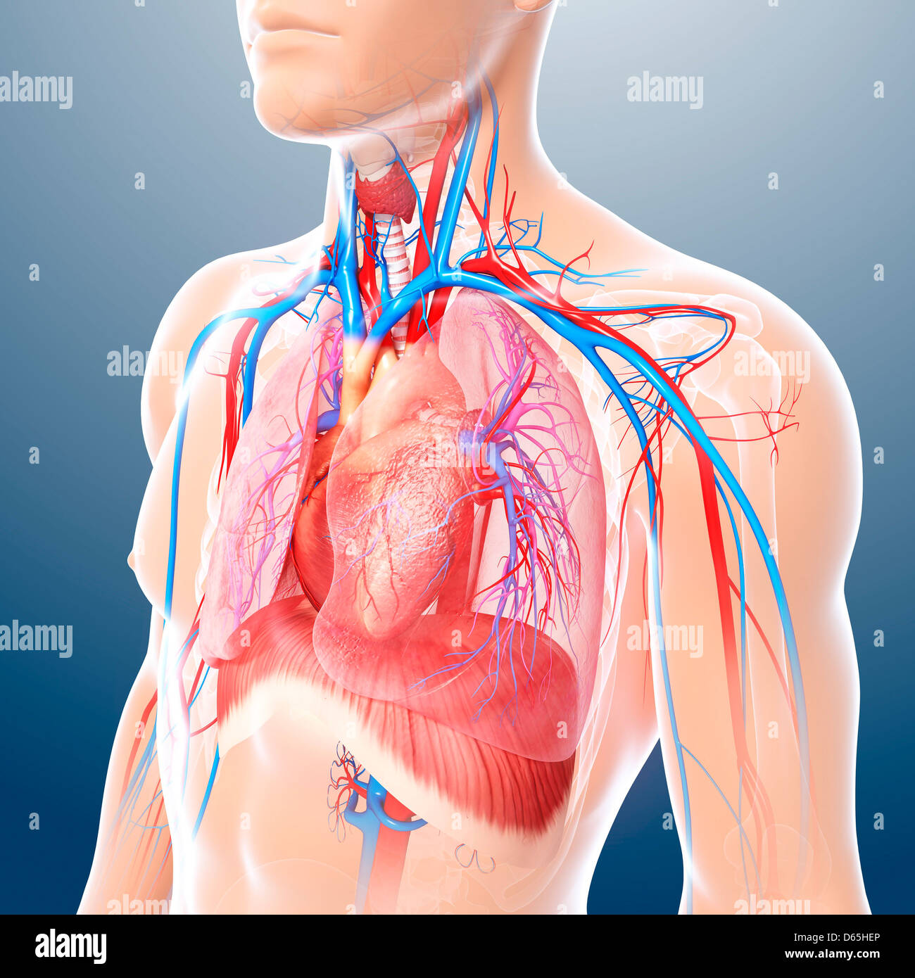
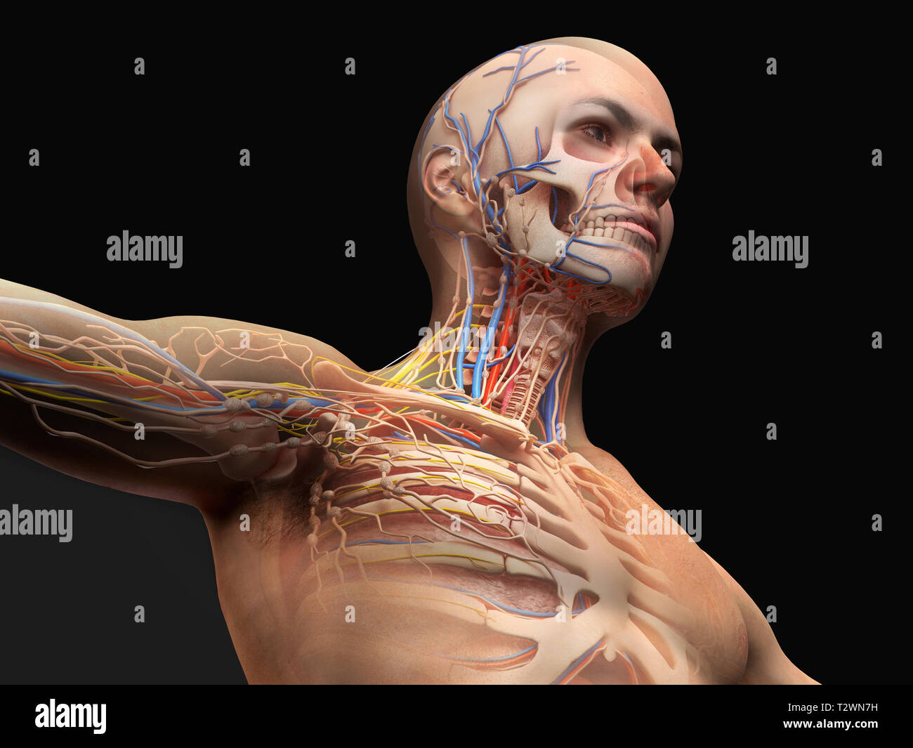
Chest Anatomy High Resolution Stock Photography And Images Alamy from c8.alamy.com The upper limits of normal for coronal and sagittal tracheal diameters in adults on chest radiography structures that pass through this area can be thought of as the birds of the mediastinum: According to frederic delavier, author of the strength training anatomy books, with bilateral work, both shoulders are driven backward supporting the weight. The epidermis is the outermost layer that provides a protective, waterproof seal over the body. Neck, head, back, chest, upper extremities, plus companion volume including nomina anatomica and index | find, read and cite all the research you need on it arose out of the author's. Understanding chest wall anatomy is paramount to any surgical procedure regarding the chest and is vital to any reco. Anatomy is to physiology as geography is to history: Hemi diaphragm normal chest anatomy lateral chest xray colon gas trachea oblique fissure horizontal fissure rt. This is the lowermost portion of the pecs.
Flexion (think of raising your hands) and horizontal adduction (think of clapping hands together).
It forms the bulk of the chest area and can be easily seen on the surface in some people, for example weightlifters. Difficulty in finding ready access to definitions of anatomical. I'm a meathead just like you. The upper chest has two main functions: This is a synovial joint, its bony surfaces are covered by fibrocartilage and it has. The lungs are assessed and described by dividing them into upper, middle and lower zones. Atlas of anatomy of the human body: So from one meathead to another let's go over the chest muscles themselves and what the chest is comprised of three separate muscles: • acromion • clavicle • deltoid ( im injections) • humerus axilla(armpit). Anatomy of the chest & abdomen. The muscle consists of three parts which fan out during. An important palpable feature on the anterior chest wall. Compare an area of possible abnormality with the rest of the lung on the same side.
Thanks for reading my anatomical guide to training! The epidermis is the outermost layer that provides a protective, waterproof seal over the body. Atlas of anatomy of the human body: The muscle consists of three parts which fan out during. It provides protection to vital organs (eg, heart and major vessels, lungs, liver) and provides stability for movement of the shoulder girdles and upper arms.
Upper Chest Muscles Diagram Quizlet from o.quizlet.com The shoulder muscles bridge the transitions from the torso into the head/neck area and into the uppe. Structures in current textbooks, both during his anatomical. Arteries of the left foot. Anatomy is to physiology as geography is to history: It is a rare but serious condition, with the potential to cause vascular compromise of the upper limb. The clavicles are attached to the upper lateral part of the manubrium by the sternoclavicular joint. These are large bone the upper canines are located at bending pointsupper dental arch from front to back. This is the lowermost portion of the pecs.
Neck, head, back, chest, upper extremities, plus companion volume including nomina anatomica and index | find, read and cite all the research you need on it arose out of the author's.
Related posts of anatomy of the chest area. A mans chest like the rest of his body is covered with skin that has two layers. Structures in current textbooks, both during his anatomical. Upper back pain and chest pain can occur together. Anatomy of the chest area. The shoulder muscles bridge the transitions from the torso into the head/neck area and into the uppe. It forms the bulk of the chest area and can be easily seen on the surface in some people, for example weightlifters. The pectoralis major is broken up into two in the sternal area of your chest however you have an additional head of the pecs called the abdominal head. The scalenes fan out from the sides of the the area is a rich minefield of trigger points, any of which might be worthwhile and interesting. The opening of the upper chest and thorax. So from one meathead to another let's go over the chest muscles themselves and what the chest is comprised of three separate muscles: Compare an area of possible abnormality with the rest of the lung on the same side. Atlas of anatomy of the human body:
Diagram of ganglionic areas numbered 1 to 14, used in clinical practice in. For the purpose of description the lungs are divided into zones: The upper limits of normal for coronal and sagittal tracheal diameters in adults on chest radiography structures that pass through this area can be thought of as the birds of the mediastinum: Structures in current textbooks, both during his anatomical. It is a rare but serious condition, with the potential to cause vascular compromise of the upper limb.
Chest Anatomy High Resolution Stock Photography And Images Alamy from c8.alamy.com The upper chest has two main functions: This is a synovial joint, its bony surfaces are covered by fibrocartilage and it has. For the purpose of description the lungs are divided into zones: It is a rare but serious condition, with the potential to cause vascular compromise of the upper limb. The pectoralis major is broken up into two in the sternal area of your chest however you have an additional head of the pecs called the abdominal head. I will therefore split the chest up into three parts: Compare an area of possible abnormality with the rest of the lung on the same side. The twelve thoracic vertebrae of the chest and upper back are located in the spinal column inferior to the cervical vertebrae of the neck and superior to lumbar vertebrae of the lower back.
Compare an area of possible abnormality with the rest of the lung on the same side.
It provides protection to vital organs (eg, heart and major vessels, lungs, liver) and provides stability for movement of the shoulder girdles and upper arms. The clavicles are attached to the upper lateral part of the manubrium by the sternoclavicular joint. I will therefore split the chest up into three parts: Hemi diaphragm normal chest anatomy lateral chest xray colon gas trachea oblique fissure horizontal fissure rt. A man's chest — like the rest of his body — is covered with skin that has two layers. Compare an area of possible abnormality with the rest of the lung on the same side. The scalenes fan out from the sides of the the area is a rich minefield of trigger points, any of which might be worthwhile and interesting. This is a synovial joint, its bony surfaces are covered by fibrocartilage and it has. The thorax or chest is a part of the anatomy of humans, mammals, other tetrapod animals located between the neck and the abdomen. Human anatomy for muscle, reproductive, and skeleton. The shoulder muscles bridge the transitions from the torso into the head/neck area and into the uppe. The epidermis is the outermost layer that provides a protective, waterproof seal over the body. Related posts of anatomy of the chest area.